Limba
 Romanian
Romanian
 English
|
Romanian
Ki67
Cat#: BSH-7302-100 100ul, BSH-7302-1 1ml, BSH-7302-RTU 7ml
Clone: BS4
S/R: human
Application: IHC
Ki67, also known as MKI67, is aprototypic cell cycle related nuclear protein, expressed by proliferating cells in all phases of the active cell cycle (G1, S, G2 and M phase). It is absent in resting (G0) cells. Ki67 antibodies are useful in establishing the cell growing fraction in neoplasms (immunohistochemically quantified by determining the number of Ki67 positive cells among the total number of resting cells = Ki67 index). In neoplastic tissues the prognostic value is comparable to the tritiated thymidine labelling index. The correlation between low Ki67 index and histologically low grade tumours is strong. Ki67 is routinely used as a neuronal marker of cell cycling and proliferation.
MBP
 |
 |
 |
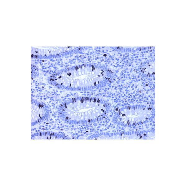
| Ki67 stained tissue sections. Ki67 Optibody (Clone: BS4) with 1:200 dilution shows same staining pattern as the optimal staining criteria of NordiQC (a). Ductal breast carcinoma shows proliferation and intensive nuclear staining pattern. Lieberkuhn crypts of colon are stained intensively with nuclear staining pattern (b, c). | ||
| Price | 2.970,00 RON (preturile sunt fara TVA) | ||||||
|---|---|---|---|---|---|---|---|
| Description |
Ki67 Cat#: BSH-7302-100 100ul, BSH-7302-1 1ml, BSH-7302-RTU 7ml Ki67, also known as MKI67, is aprototypic cell cycle related nuclear protein, expressed by proliferating cells in all phases of the active cell cycle (G1, S, G2 and M phase). It is absent in resting (G0) cells. Ki67 antibodies are useful in establishing the cell growing fraction in neoplasms (immunohistochemically quantified by determining the number of Ki67 positive cells among the total number of resting cells = Ki67 index). In neoplastic tissues the prognostic value is comparable to the tritiated thymidine labelling index. The correlation between low Ki67 index and histologically low grade tumours is strong. Ki67 is routinely used as a neuronal marker of cell cycling and proliferation.
|
||||||