Limba
 Romanian
Romanian
 English
|
Romanian
CEA
Cat#: BSH-7437-100 100ul, BSH-7437-1 1ml, BSH-7437-RTU 7ml
Clone: BS33
S/R: human
Application: IHC
Tissue control: Appendix, colon and liver (negative)
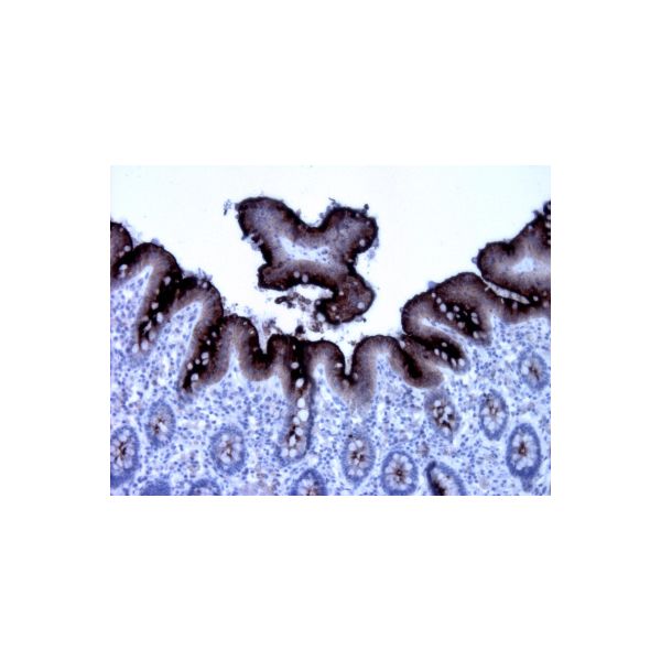
CEA are useful in identifying the origin of various metastatic adenocarcinomas and in distinguishing pulmonary adenocarcinomas (60 to 70% are CEA+) from pleural mesotheliomas (rarely or weakly CEA+).The carcinoembryonic antigen (CEA) is a member of a large family of glycoproteins and a useful tumor marker for adenocarcinoma. Tissue specificity: Found in adenocarcinomas of endodermally derived digestive system epithelium and fetal colon.
 |
 |
 |
| CEA stained tissue sections. CEA optibody (Clone: BS33) staining with 1:250 dilution is intensive and specific (a, b, c) without staining of the liver bile ducts (negative control) (c). The signal to noise ratio is high. Luminal part of columnar epithelia stained strongly (appendix, b, c) and liver stained negatively (c). Colorectal cancer metastase in lymph node stained strongly with CEA optibody | ||
| Price | 2.970,00 RON (preturile sunt fara TVA) | ||||||
|---|---|---|---|---|---|---|---|
| Description |
CEA Cat#: BSH-7437-100 100ul, BSH-7437-1 1ml, BSH-7437-RTU 7ml CEA are useful in identifying the origin of various metastatic adenocarcinomas and in distinguishing pulmonary adenocarcinomas (60 to 70% are CEA+) from pleural mesotheliomas (rarely or weakly CEA+).The carcinoembryonic antigen (CEA) is a member of a large family of glycoproteins and a useful tumor marker for adenocarcinoma. Tissue specificity: Found in adenocarcinomas of endodermally derived digestive system epithelium and fetal colon.
|
||||||