The store will not work correctly in the case when cookies are disabled.

83601, µ-Slide VI 0.4 µ-Pattern RGD, sqr20, pit110, hex:
#1.5 polymer coverslip, micropatterned surface with RGD motif, 20 μm squares, 110 μm pitch, hexagonal layout, surface passivation with Bioinert, sterilized, 10 Pcs/box

83802, µ-Slide 8 Well high µ-Pattern RGD, cir100, pit500, hex: #1.5 polymer coverslip, micropatterned surface with RGD motif, 100 μm circles, 500 μm pitch, hexagonal layout, surface passivation with Bioinert, sterilized, 10 Pcs/box
83601-S, µ-Slide VI 0.4 µ-Pattern RGD, sqr20, pit110, hex Trial Pack: #1.5 polymer coverslip, micropatterned surface with RGD motif, 20 μm squares, 110 μm pitch, hexagonal layout, surface passivation with Bioinert, sterilized, 2 Pcs/box
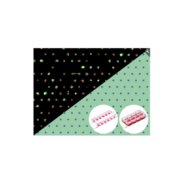
A micropatterned surface for single cell assays with fluorescence microscopy readout
A micropatterned surface for single cell assays with fluorescence microscopy readout
- Two different single-cell patterns with ideal spacing for various single cell assays
- Easy handling without preparation: Unpack and start
- Excellent optical-quality imaging chamber for high-resolution microscopy
Specifications
|
µ-Slide geometry
|
See product page
µ-Slide 8 Well high or µ-Slide VI 0.4
|
|
Binding motif
|
RGD
|
|
Pattern shape
|
Square
|
|
Side length
|
20 μm or 30 µm
|
|
Pitch
|
110 μm
|
|
Pattern layout
|
Hexagonal
|
|
Surface passivation
|
Bioinert
|
|
Number of patterns
- µ-Slide 8 Well high
- µ-Slide VI 0.4
|
ca. 9,500 per well
ca. 6,000 per channel
|
Technical Features
- µ-Slide that has a micropatterned surface with a covalently bound RGD motif (binding motif from fibronectin) on the ibidi Bioinert surface
- Due to the Bioinert surface, cells can attach only on the patterned area, even during long-term cultivation for days or weeks
- Bioinert surface passivation—superior to standard ultra-low attachment (ULA) surfaces:
- No cell or protein adhesion
- Long-term stability
- Biologically inert
- Bioinert is layered onto the ibidi Polymer Coverslip, which provides the highest optical quality for imaging when using high-resolution microscopy
- Non-fluorescent patterns, not visible in phase contrast microscopy
- Patterns are printed on either the µ-Slide 8 Well high or µ-Slide VI 0.4
- Compatible with staining and fixation solutions
- Fully biocompatible materials
Mai multe informatii
| Price |
792,00 RON (preturile sunt fara TVA) |
| Description |
A micropatterned surface for single cell assays with fluorescence microscopy readout
- Two different single-cell patterns with ideal spacing for various single cell assays
- Easy handling without preparation: Unpack and start
- Excellent optical-quality imaging chamber for high-resolution microscopy
Specifications
|
µ-Slide geometry
|
See product page
µ-Slide 8 Well high or µ-Slide VI 0.4
|
|
Binding motif
|
RGD
|
|
Pattern shape
|
Square
|
|
Side length
|
20 μm or 30 µm
|
|
Pitch
|
110 μm
|
|
Pattern layout
|
Hexagonal
|
|
Surface passivation
|
Bioinert
|
|
Number of patterns
- µ-Slide 8 Well high
- µ-Slide VI 0.4
|
ca. 9,500 per well
ca. 6,000 per channel
|
Technical Features
- µ-Slide that has a micropatterned surface with a covalently bound RGD motif (binding motif from fibronectin) on the ibidi Bioinert surface
- Due to the Bioinert surface, cells can attach only on the patterned area, even during long-term cultivation for days or weeks
- Bioinert surface passivation—superior to standard ultra-low attachment (ULA) surfaces:
- No cell or protein adhesion
- Long-term stability
- Biologically inert
- Bioinert is layered onto the ibidi Polymer Coverslip, which provides the highest optical quality for imaging when using high-resolution microscopy
- Non-fluorescent patterns, not visible in phase contrast microscopy
- Patterns are printed on either the µ-Slide 8 Well high or µ-Slide VI 0.4
- Compatible with staining and fixation solutions
- Fully biocompatible materials
|
 Romanian
 English